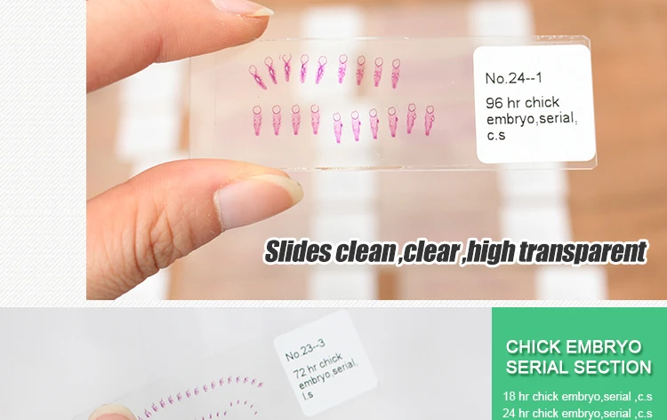
96 Hour Chick Embryo Serial Section

Section, 24 hr sagital, 33 hr serial x-sections, 48 or 56 hr. REFERENCE: Patten, Embryology of the chick. Arteries and veins) in the 96 hr whole mount. 4 Day (96 hour) Chick Embryo. Extending out from the embryo is a thin delicate membrane with blood vessels at its surface. This membrane, called the allantois, expands as the embryo develops until it completely lines the shell.
Chick Embryo 96 Hours Serial Sag Section Prepared Microscope Slide Limited Availability of Chick Embryo 96 Hours Serial Sag Section Prepared Microscope Slide EE12-3 Chick Embryo 96 Hours Serial Sag Section Prepared Microscope Slide 96 hr chick; serial sagital section A 10% discount applies if you order more than 10 of this item and 15% discount applies if you order more than 25 of this item. Triarch Incorporated offers superior prepared microscope slides. While we produce over 2300 different slides, we also make, and slides. In addition, we offer affordable quality microscopes from and for less. Use coupon code SWIFT10 for an additional 10% off our already low Swift prices. Educational digital images are also available for purchase at high resolution magnifications (10x, 25x, and 100x). CS = Cross Section: So the slide shows a thin section through the transverse plane of an organism.
LS = Longitudinal Section: So the slide shows a vertical section of the organism along the longest plane. WM = Whole Mount: So the slide shows an entire organism or structure, as indicated, is preserved on the slide CRT = Cross Section, Radial Section, and Tangential Section: So the slide shows sections of wood along the transverse, radial, and tangential planes. Sag = Sagittal Section: So the slide shows a thin section through the sagittal plane through the midline. Serial Sections = So the slide shows consecutive sections of the organism. Rep = Representative Sections (Embryology): So the slide shows one section of the organism from each typical area of study. Psicopatologia uma abordagem integrada pdf to doc document. Triarch Incorporated’s name is based on a botanical slide that illustrates three ridges of xylem found in the vascular cylinder of the Ranunculus root. Our founder, George H.
Conant, Ph.D., had three principles in mind: Accuracy, Service, and Dependability. He incorporated these into the Triarch logo based on the triarch vascular cylinder.

This membrane is made up of a bladderlike median ventral diverticulum of the hindgut endoderm, covered with splanchnic mesoderm. It connects with the hindgut, which will be found only in sections posterior to the posterior intestinal portal at this stage of development. Ultimately, this double membrane will fill the exocoel, and its outer layer of mesoderm will fuse with mesoderm of the chorion and the aminion and finally with the splanchnic mesoderm of the yolk sac splanchnopleure. Its function in the chick is related to respiration and excretion. The entire outer covering of the chick embryo is of ectodermal origin and is made up largely of squamous epithelium but will later also include horny scales, feather germs, quills and barbs, claws, beak coverings, and a temporary eggtooth. By evaginations from the surface, the linings of the following structures are also derived from ectoderm: the mouth (stomodeal portion and stomodeal hypophysis); cloaca (proctodeal portion); visceral clefts (peripheral halves); nostrils; eye chamber and lens; otic vesicles; and external auditory meatus.